
Blood Collection Tubes and Anticoagulants: Practical Revision + 15 MCQs
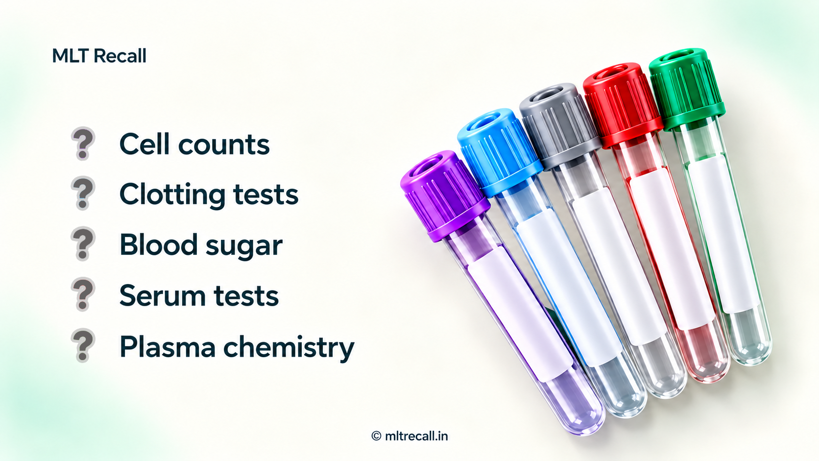
MLT Recall | DMLT/BMLT revision for AIIMS CRE and ESIC Lab Technician preparation
A patient comes to the collection counter with a request for CBC, fasting blood sugar, LFT and PT/INR. All four tests need blood, but they do not all need the same specimen.
This is where a lab technician's basic knowledge matters: selecting the correct container, collecting the required quantity, mixing properly and recognising an unsuitable sample before testing starts.
Learn this topic in a simple sequence:
Test requested → specimen required → additive needed → collection precautions.
The questions below are original practice questions for recruitment preparation, not previous-year questions or an official AIIMS/ESIC protocol. For actual collection, follow your laboratory's SOP and the tube label. Cap colours mentioned here are common examples; they can differ between products.
1. Which tube will you pick at the collection counter?
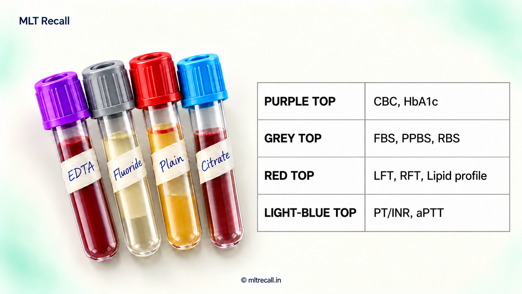
For the four investigations above, the usual arrangement is:
- CBC: EDTA tube — anticoagulated whole blood.
- Fasting blood sugar: fluoride-containing glucose tube — plasma after processing.
- LFT: plain or clot-activator serum tube — allow clotting, then separate serum.
- PT/INR: sodium citrate tube — citrated plasma after processing.
The important point is the additive and specimen, not simply remembering “purple, grey, red and blue”. A correctly coloured tube with the wrong additive or inadequate filling is still unsuitable.
2. Main revision table: what is the difference?
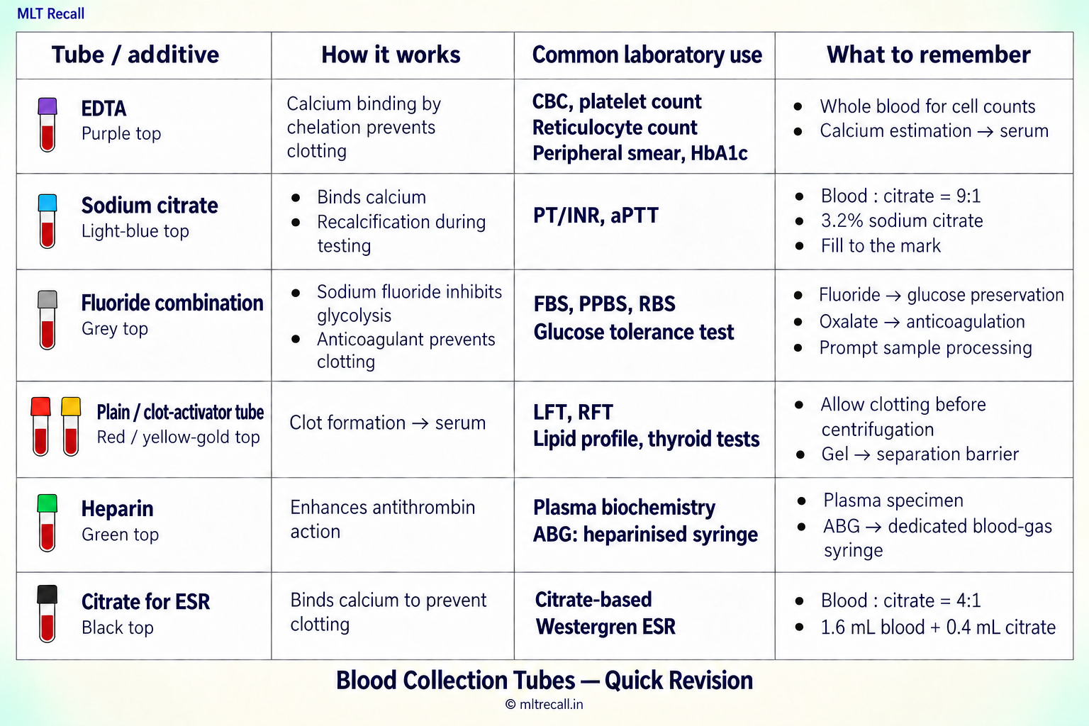
Read every ratio as blood : anticoagulant. Serum is common for routine biochemistry, but some methods accept or require plasma. Select the specimen required by the test method.
3. EDTA: the routine haematology anticoagulant
EDTA stands for ethylenediaminetetraacetic acid. It binds calcium, which is needed for coagulation, and keeps the blood from clotting.
In routine haematology, EDTA is used because blood cells can be counted and examined in an anticoagulated sample. Prepare the peripheral smear promptly; prolonged storage can alter cell morphology.
Remember these practical points:
- Mix the sample gently by inversion soon after collection. Use the inversion count on the product instructions.
- Collect the specified quantity. Too little blood leaves excess EDTA relative to the sample and may alter cell size and indices.
- Check for clots before running a doubtful sample on the analyser.
- A clotted CBC sample needs recollection. Removing the clot does not restore the cells trapped in it.
Common confusion: blood sugar versus HbA1c. Both are used in diabetes assessment, but routine collection differs. A fluoride tube is commonly used for blood glucose; EDTA whole blood is commonly used for HbA1c. HbA1c measures glycated haemoglobin in red cells, so separated serum is unsuitable.
Another important point: potassium EDTA contamination can produce falsely high potassium and low calcium. Investigate an unexpected result pattern; do not assume every high potassium value is a collection error.
4. Sodium citrate: remember the purpose and the ratio
PT/INR and aPTT: 9:1
Routine coagulation collection uses nine parts blood and one part sodium citrate.
For example:
2.7 mL blood + 0.3 mL citrate = 3.0 mL total mixture.
The ratio refers to the amount of blood compared with citrate, not the total mixture compared with citrate. Commercial tubes already contain the additive: collect to the specified fill mark rather than preparing the mixture yourself.
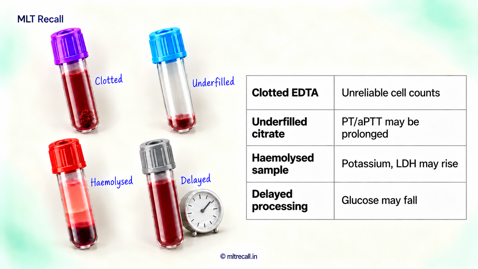
An underfilled citrate tube has too much citrate for the amount of blood collected. It can give falsely prolonged PT/aPTT. Do not report it as a patient abnormality without checking sample suitability. Do not top it up with blood from an EDTA tube.
Conventional citrate-based Westergren ESR: 4:1
The familiar teaching ratio is four parts blood to one part citrate.
1.6 mL blood + 0.4 mL citrate = 2.0 mL total mixture.
Traditional teaching commonly mentions 3.8% citrate for Westergren ESR; commercial ESR systems may use other specified formulations, including 3.2%. Follow the method being used.
For examination revision, distinguish:
- Citrate-based Westergren: 4:1 blood:citrate.
- Wintrobe: undiluted anticoagulated blood, commonly EDTA blood; double oxalate is also described in classical teaching.
- Automated ESR: the instrument's specified sample, often EDTA whole blood.
Therefore, “ESR always needs a separate citrate tube” is incorrect.
5. Fluoride tube: useful, but processing still matters
A common OPD request is FBS, PPBS or RBS. A grey fluoride-containing tube is routinely associated with these tests.
In the familiar sodium fluoride–potassium oxalate combination:
- Sodium fluoride: inhibits the glycolytic enzyme enolase.
- Potassium oxalate: prevents coagulation by precipitating calcium.
Do not interchange their functions in an MCQ.
Blood cells continue to consume glucose after collection. If processing is delayed, glucose can fall. Fluoride does not immediately stop all glycolysis, so prompt transport and processing remain important.
For fasting and post-meal samples, record the collection time and identify the sample correctly. Two identical grey tubes without clear fasting/post-meal labels can still lead to a wrong report.
6. Serum, plasma and whole blood: three different specimens
Whole blood: cells remain mixed with the liquid component. Example: EDTA blood for CBC or HbA1c. Do not centrifuge a CBC sample to obtain serum.
Plasma: liquid separated from anticoagulated blood. It contains fibrinogen. Example: citrated plasma for PT/aPTT.
Serum: liquid separated after blood has clotted. Fibrinogen is used up during clot formation. Example: serum for many routine LFT and thyroid assays.
Exam recall: plasma has fibrinogen; serum normally lacks fibrinogen. This does not mean that every coagulation factor is absent from serum.
A serum sample should be allowed to clot properly before centrifugation. Incomplete clotting can leave fibrin strands that interfere with pipetting or analyser sampling. Do not remove an unexpected clot from a supposed plasma specimen and relabel the liquid as serum.
7. Common sample problems you should recognise
Clot in an EDTA sample: cell counts may be unreliable because cells are trapped in the clot. Recollect rather than picking out the clot and running the remaining blood.
Pink or red serum/plasma: consider haemolysis. Haemolysis can falsely increase potassium and LDH and interfere with other tests. Suitability depends on the degree of haemolysis and the assay; follow the rejection criteria.
Sample from an arm receiving an IV infusion: infusion fluid may contaminate or dilute the sample. Prefer the other arm when possible. Dextrose contamination can falsely increase glucose.
Prolonged tourniquet application or repeated fist pumping: can alter results. Avoid leaving the tourniquet on unnecessarily and do not ask the patient to repeatedly pump the fist.
Poor mixing: can allow small clots to form. Vigorous shaking: can damage cells. Mix by gentle inversion as instructed.
Wrong label or patient mismatch: stop processing and resolve the identity problem under the SOP. A technically perfect specimen cannot give a useful report if it belongs to the wrong patient.
8. Fifteen practice MCQs
Choose one best answer. The questions combine basic facts, routine bench situations and common recruitment-exam distinctions.
1. An OPD patient needs CBC, fasting blood glucose and LFT. Which combination is most appropriate for routine collection?
A. Citrate for CBC, EDTA for glucose, serum for LFT
B. EDTA for CBC, fluoride-containing tube for glucose, serum tube for LFT
C. Serum for CBC, citrate for glucose, EDTA for LFT
D. Fluoride-containing tubes for all three tests
Show answer and explanation
Answer: B. CBC needs anticoagulated whole blood; fluoride tubes are commonly used for glucose; serum is commonly used for LFT. One collection tube cannot automatically serve every test on a request form.
2. A patient brings a request for HbA1c. Which specimen is commonly required?
A. Citrated plasma
B. Serum from a plain tube
C. Fluoride plasma
D. EDTA whole blood
Show answer and explanation
Answer: D. HbA1c is measured in haemoglobin within red cells. Remember the distinction: glucose — commonly fluoride plasma; HbA1c — commonly EDTA whole blood.
3. Blood for serum calcium estimation is accidentally collected into an EDTA tube. Why is this unsuitable?
A. EDTA binds calcium
B. EDTA adds calcium to the specimen
C. EDTA acts by diluting blood in a 9:1 ratio
D. EDTA forms a separator-gel barrier around calcium
Show answer and explanation
Answer: A. EDTA chelates calcium. This is how it prevents clotting and why an EDTA specimen is unsuitable for routine calcium estimation. Collect the appropriate specimen again.
4. In a sodium fluoride–potassium oxalate tube, which pairing is correct?
A. Fluoride — anticoagulant; oxalate — clot activator
B. Fluoride — separator; oxalate — preservative
C. Fluoride — antiglycolytic agent; oxalate — anticoagulant
D. Fluoride — clot activator; oxalate — antiglycolytic agent
Show answer and explanation
Answer: C. Fluoride inhibits enolase in glycolysis. Oxalate precipitates calcium and prevents clotting. Their functions are a common source of confusion.
5. A small clot is found in an EDTA sample sent for platelet count. What should the technician do?
A. Remove the clot and report the remaining sample
B. Request recollection according to the rejection procedure
C. Add saline and multiply the result by two
D. Transfer the blood into a fresh EDTA tube
Show answer and explanation
Answer: B. Platelets and other cells may already be trapped in the clot. Neither removing it nor changing the container restores the original cell count.
6. A light-blue citrate tube for PT/INR contains much less blood than its fill mark. What problem can this cause?
A. Falsely shortened PT because citrate is insufficient
B. No change in PT because the additive is still citrate
C. Clot formation because an underfilled tube has too little citrate relative to blood
D. Falsely prolonged PT because citrate is excessive relative to blood
Show answer and explanation
Answer: D. Underfilling disturbs the blood:citrate ratio. Excess citrate can affect the calcium-dependent clotting test. Check specimen acceptance before testing.
7. A coagulation tube contains 0.3 mL citrate. How much blood is needed for a 9:1 blood:citrate ratio?
A. 2.7 mL
B. 3.0 mL
C. 1.2 mL
D. 0.9 mL
Show answer and explanation
Answer: A. Blood volume = 9 × 0.3 = 2.7 mL. The final mixture is 3.0 mL. Do not confuse blood volume with total volume.
8. For a conventional citrate-based Westergren preparation, 0.4 mL citrate is used. Which blood volume gives the correct ratio?
A. 3.6 mL
B. 2.0 mL
C. 1.6 mL
D. 0.4 mL
Show answer and explanation
Answer: C. Blood:citrate is 4:1, so 4 × 0.4 = 1.6 mL blood. A 3.6 mL blood volume would give 9:1, the coagulation ratio.
9. After centrifugation, the liquid above the cells in a properly collected anticoagulated sample is:
A. Serum, containing fibrinogen
B. Plasma, containing fibrinogen
C. Serum, without fibrinogen
D. Plasma, without fibrinogen
Show answer and explanation
Answer: B. Anticoagulation prevents clot formation, so the separated liquid is plasma and retains fibrinogen. Serum is separated after clotting.
10. An ABG sample is requested from the emergency department. Which collection device is appropriate?
A. Plain serum tube
B. Routine citrate coagulation tube
C. EDTA tube used for CBC
D. Dedicated heparinised blood-gas syringe
Show answer and explanation
Answer: D. ABG samples are collected using the specified heparinised blood-gas syringe. Avoid air bubbles and send promptly according to the blood-gas procedure.
11. A serum sample is visibly haemolysed. Which pair of results may be falsely increased because of red-cell breakdown?
A. Potassium and LDH
B. Sodium and chloride
C. Glucose and bicarbonate
D. Calcium and albumin
Show answer and explanation
Answer: A. Red cells contain potassium and LDH. Their release can increase measured values. Assess haemolysis and the affected tests before releasing results.
12. A blood glucose sample is collected close to an IV line running dextrose. The glucose result is unexpectedly very high. Which collection error should be considered?
A. Glycolysis during transport
B. Incomplete clotting in a serum tube
C. Contamination by the dextrose infusion
D. Underfilling of a citrate tube
Show answer and explanation
Answer: C. Dextrose contamination can falsely increase glucose. Delayed processing with ongoing glycolysis generally lowers glucose, so it does not explain this collection-related rise.
13. Which statement correctly distinguishes conventional Wintrobe and citrate-based Westergren ESR methods?
A. Both always use a 9:1 blood:citrate mixture
B. Wintrobe uses undiluted anticoagulated blood; citrate-based Westergren uses a 4:1 mixture
C. Wintrobe uses serum; Westergren uses plasma without cells
D. Both require fluoride–oxalate blood
Show answer and explanation
Answer: B. Wintrobe uses undiluted anticoagulated blood. In the conventional citrate-based Westergren preparation, the blood:citrate ratio is 4:1. Automated ESR systems have their own sample requirements.
14. A blood sample for glucose remains unprocessed for several hours. Which statement is most accurate?
A. Glucose must rise because red cells release glucose
B. A grey cap guarantees that glucose never changes
C. Fluoride replaces the need for timely transport
D. Glucose may fall because cells continue glycolysis, particularly before fluoride becomes fully effective
Show answer and explanation
Answer: D. Fluoride does not immediately stop all glycolysis. A correct tube still needs timely transport and processing.
15. All of the following are appropriate collection practices EXCEPT:
A. Filling an underfilled citrate tube with blood taken from an EDTA tube
B. Matching the patient details on the tube and request
C. Gently inverting an additive tube as instructed
D. Allowing a serum specimen to clot properly before centrifugation
Show answer and explanation
Answer: A. EDTA contaminates the citrate specimen and makes it unsuitable for routine coagulation testing. Do not transfer blood between additive tubes to correct a collection mistake.
9. One-minute revision
- CBC and HbA1c: commonly EDTA whole blood.
- FBS, PPBS and RBS: commonly fluoride-containing tube.
- PT/INR and aPTT: citrate plasma; 9:1 blood:citrate.
- Conventional citrate-based Westergren ESR: 4:1 blood:citrate.
- Wintrobe ESR: undiluted anticoagulated blood.
- ABG: dedicated heparinised blood-gas syringe.
- Plasma contains fibrinogen; serum normally lacks it.
- Clotted CBC sample: recollect; do not remove the clot and report.
- Underfilled citrate: clotting times may be falsely prolonged.
- Haemolysis: potassium and LDH may rise; delayed glucose processing can lower glucose.
Remember: the correct report begins with the correct patient, correct tube and correct sample.